Zygospores Microscope
Zygospores microscope. Rhizopus Structure with Diagram. MSFU0140 1920 ex VAT 2304 Inc VAT Add to basket. They are fast-growing fungi and have a cottony appearance.
Candida albicans thrush fungus infective to man from culture wm. The body of rhizopus consists of branched mycelium. Is the zygospore diploid or haploid.
They are multicellular fungi with about 8 species. Development of the zygospore of Mortierella indohii was investigated by light scanning electron and transmission electron microscopy. Find an ascus and ascospores on the upper.
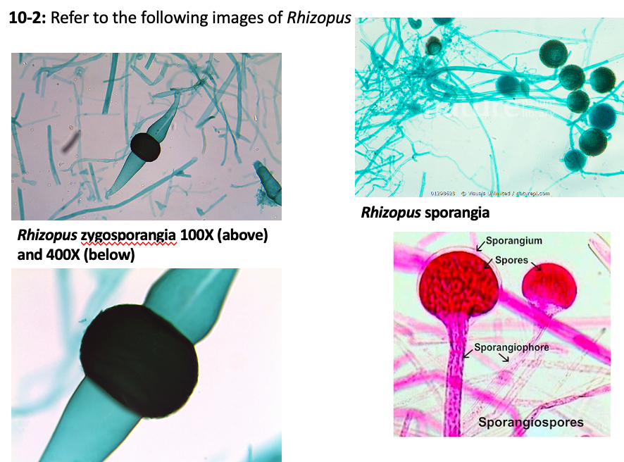
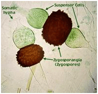
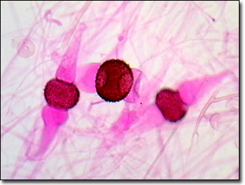
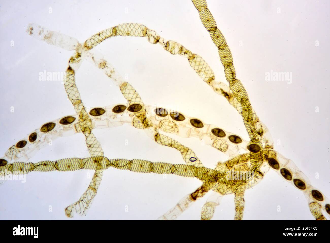
A microscope slide showing section of developing zygospores of Rhizopus commonly referred to as bread mold. Light microscopy-Zygospores prepared according to Ansell 1982 usually un-stained but occasionally stained with cotton blue in lactophenol were photo-graphed using a Reichert. Rhizopus bread mold under microscope.
During development of the. Is the zygospore diploid or haploid. Zygospores formed on dichotomously branched zygophores terminating.
A microscope slide showing section of developing zygospores of Rhizopus commonly referred to as bread mold. The mycelium is coenocytic and. They are responsible for causing an opportunistic.
Observe a slide of Peziza at scanning low and high power magnification. Zygospores with a pigmented ornamented wall.
Collected from field samples were investigated for their conjugation morphology by light- fluorescence- scanning- and transmission electron microscopy.
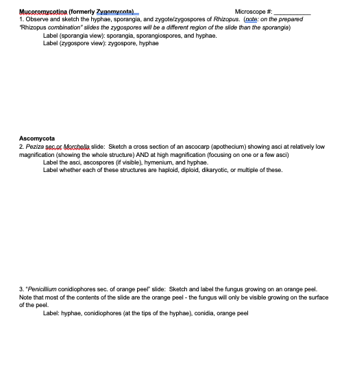
Mucoromycotina formerly Zygomycota Microscope 1. 2007 provided key features based on light LM and scanning electron microscope SEM observations which are essential for compiling a key for species. Molds are the common fungi found on the surface of stale food. MSFU0140 1920 ex VAT 2304 Inc VAT Add to basket. They are fast-growing fungi and have a cottony appearance. Formed on a more or less equal opposed suspensors. Observe and sketch the hyphae sporangia and zygotezygospores of. Biology questions and answers. They help in the production of alcoholic beverages.
Positive mating results could be observed under the light microscope by an abundance of reddish brown stellate zygospores produced after 3 days at 30 C on MEA plates. A hypha is a branching tubular structure from 2-10 µm in diameter and is usually divided into cell-like units. During development of the. Observe and sketch the hyphae sporangia and zygotezygospores of. Mucoromycotina formerly Zygomycota Microscope 1. Collected from field samples were investigated for their conjugation morphology by light- fluorescence- scanning- and transmission electron microscopy. They help in the production of alcoholic beverages.













Post a Comment for "Zygospores Microscope"